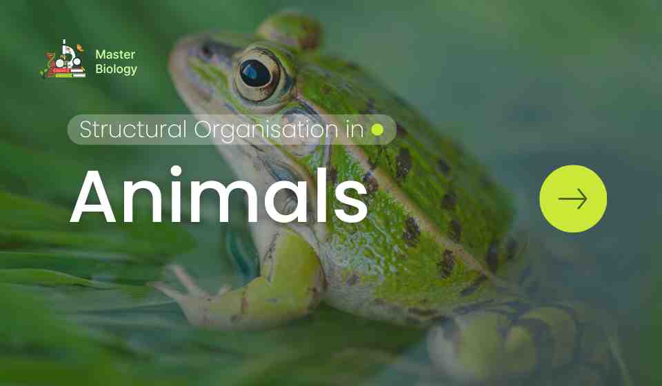
Structural Organisation in Animals reveals how the animal kingdom showcases a breathtaking spectrum of complexity, from microscopic sponges to colossal blue whales. This complexity arises not from new materials, but from the hierarchical and integrated organisation of simple biological units. This article explores this structural organisation, beginning with the foundational level of animal tissues, and then uses the common Indian bullfrog (Rana tigrina) as a model organism to illustrate how these tissues combine into organs and systems that sustain life.
I. Animal Tissues: The Building Blocks
Tissues are groups of cells similar in structure and function, along with their extracellular matrix. They are classified into four primary types:
1. Epithelial Tissue:
- Structure: Tightly packed cells forming continuous sheets, with little intercellular matrix. Rest on a basement membrane. Can be simple (single layer) or stratified (multiple layers). Shapes include squamous (flat), cuboidal (cube-like), and columnar (pillar-like).
- Function: Protection (skin), absorption (intestinal lining), secretion (glands), and sensory reception (taste buds).
2. Connective Tissue:
- Structure: Characterized by loosely spaced cells embedded in a non-living, often fibrous, intercellular matrix. Includes the most diverse tissue types.
- Function:
- Areolar: Fills space between organs, provides cushioning.
- Adipose: Stores fat, provides insulation.
- Skeletal: Bone (rigid, supportive) and Cartilage (flexible, supportive).
- Vascular: Blood (fluid matrix: plasma) for transport; Lymph for immunity and drainage.
3. Muscular Tissue:
- Structure: Composed of elongated, excitable cells (muscle fibres) containing contractile proteins (actin and myosin).
- Function: Movement.
- Skeletal (Striated): Voluntary, attached to bones.
- Smooth (Non-striated): Involuntary, found in visceral organs.
- Cardiac: Involuntary, striated, branched fibers found only in the heart.
4. Nervous Tissue:
- Structure: Consists of neurons (excitable cells with dendrites, a cell body, and a long axon) and neuroglial cells (support and protect neurons).
- Function: Perception of stimuli, integration of information, and conduction of electrochemical impulses to coordinate body functions.
These tissues do not work in isolation. They combine to form organs (e.g., the stomach contains all four tissue types), which further integrate into organ systems.
II. The Frog: A Model for Vertebrate Systems
The frog, an amphibian, is an excellent model to study vertebrate anatomy due to its intermediate position in evolution and relative structural simplicity. Its morphology (external form) includes a streamlined body divided into head and trunk, paired limbs (hind limbs adapted for jumping and swimming), and moist, scale-less skin.
Its anatomy (internal structure) reveals how organ systems are assembled from tissues. Below is an exploration of key systems:
1. Digestive System
- Morphology & Anatomy: The alimentary canal is a short, coiled tube from the mouth to the cloaca. Key structures include a mouth with a sticky tongue, oesophagus, bag-like stomach, long, coiled small intestine (duodenum and ileum), short large intestine, cloaca, and digestive glands (liver with a gallbladder, pancreas).
- Function: Digestion is both extracellular (in the stomach and intestine via enzyme action) and intracellular (in certain cells). The stomach stores and churns food with gastric juices. The small intestine is the primary site for enzymatic digestion and nutrient absorption. The liver produces bile for fat emulsification, and the pancreas secretes digestive enzymes into the duodenum.
2. Circulatory System
- Morphology & Anatomy: Frogs have a closed circulatory system with a three-chambered heart (two atria, one ventricle), blood vessels, and blood (connective tissue). A unique venous connection, the hepatic portal system, carries blood from the gut to the liver.
- Function: The system transports gases, nutrients, hormones, and wastes. Oxygenated and deoxygenated blood mix partially in the single ventricle. The double circulation consists of:
- Pulmonary Circulation: Heart → Lungs → Heart (for oxygenation).
- Systemic Circulation: Heart → Body → Heart (for distributing oxygenated blood).
- Cutaneous Circulation: Skin acts as a secondary respiratory organ, with rich blood supply for gas exchange.
3. Respiratory System
- Morphology & Anatomy: Frogs exhibit multiple modes of respiration. They possess paired, sac-like lungs with simple internal folds. The skin is thin, moist, and highly vascularized. The bucco-pharyngeal cavity lining is also vascular.
- Function:
- Cutaneous Respiration: Dominant during hibernation (aestivation) and in water. Oxygen diffuses directly through the moist skin into blood capillaries.
- Buccopharyngeal Respiration: On land, gas exchange occurs through the lining of the mouth cavity.
- Pulmonary Respiration: During increased activity, air is drawn into the lungs via positive pressure breathing (forcing air in by lowering the mouth floor). Lungs provide a larger surface area for gas exchange.
4. Nervous System
- Morphology & Anatomy: Divided into:
- Central Nervous System (CNS): Brain (encased in cranium: forebrain, midbrain, hindbrain) and spinal cord.
- Peripheral Nervous System (PNS): Cranial nerves (10 pairs) and spinal nerves.
- Autonomic Nervous System (ANS): Sympathetic and parasympathetic chains.
- Function: The brain is the central control unit. The medulla oblongata (hindbrain) controls vital functions like respiration and heartbeat. The olfactory lobes (forebrain) are well-developed for smell. The optic lobes (midbrain) process vision. The spinal cord relays signals and mediates reflex actions. The ANS regulates involuntary functions of visceral organs.
5. Reproductive System
- Morphology & Anatomy:
- Male: A pair of yellowish, ovoid testes attached to kidneys. Sperms travel through vasa efferentia to kidneys and exit via ureters, which thus act as urinogenital ducts, opening into the cloaca.
- Female: A pair of large, lobed ovaries with a functional oviduct on each side, separate from the kidneys. The oviducts open into the cloaca independently.
- Function: Reproduction is sexual. Fertilization is external in water. During amplexus (mating embrace), the female releases eggs, and the male releases sperm over them. The fertilized eggs undergo external development, often through a larval stage (tadpole), showcasing metamorphosis.
Integration: The Symphony of Life
The frog exemplifies how the hierarchical structural organisation—from tissues to systems—culminates in a functioning organism. Its digestive system provides nutrients, absorbed into the circulatory system, which is powered by the heart. The respiratory system (lungs, skin, mouth) supplies oxygen to the blood and removes carbon dioxide, a process coordinated by the nervous system. This same nervous system, along with hormonal signals, regulates the reproductive system to ensure species survival.
This integration is only possible because specialized tissues form organs designed for specific tasks, and these organs communicate via circulatory and neural pathways. Studying the frog provides a fundamental blueprint for understanding the structural and functional principles that underlie all complex animal life, including our own. It is a testament to the elegance and efficiency of biological organisation.